Figure 4.

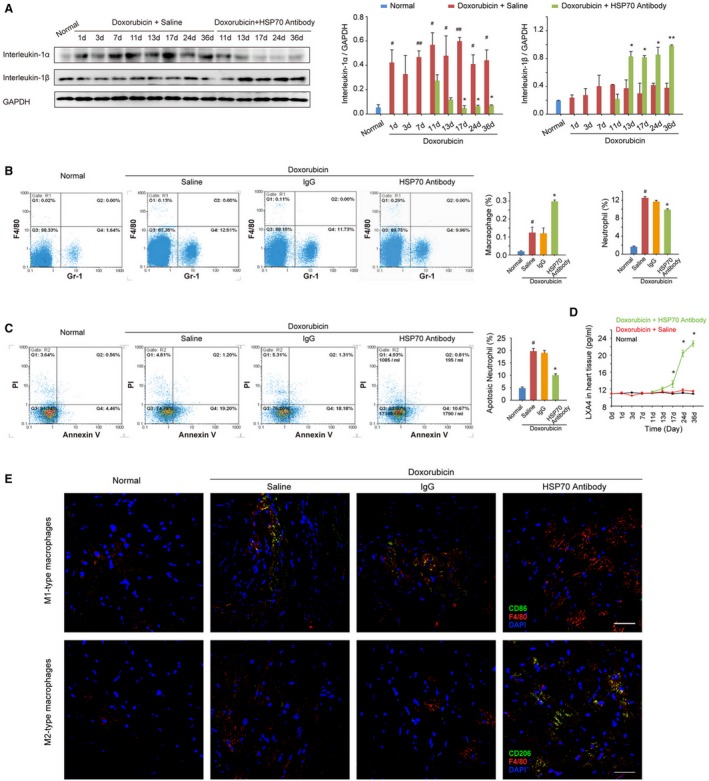
Antagonism of extracellular HSP (heat shock protein) 70 promotes resolution of inflammation in the heart tissue of doxorubicin‐treated mice. A, The time‐dependent expressions of interleukin‐1α and interleukin‐1β in the mouse myocardium after administration of doxorubicin and/or HSP70 antibody. Representative immunoblots and the ratio of the indicated protein to GAPDH are presented. B, The heart single‐cell suspensions were prepared, and the Gr‐1–positive neutrophils and F4/80‐positive macrophages were determined by flow cytometry analysis. Representative scattergrams and the percentage of neutrophils and macrophages are presented. C, The apoptosis of neutrophils was determined by the annexin V and propidium iodide (PI) staining, followed by flow cytometry. Representative scattergrams and percentage of apoptotic neutrophils are presented. D, At the indicated time points, the contents of lipoxin A4 (LXA4) in heart tissue homogenates were detected by ELISA analysis. All data are the mean±SEM of 3 assays. # P<0.05, ## P<0.01, compared with normal mice; *P<0.05, **P<0.01, compared with doxorubicin‐treated mice. E, Recruitment and infiltration of M1‐type macrophages (F4/80+CD86+) and M2‐type macrophages (F4/80+CD206+) in myocardium were detected by confocal microscopy analysis on day 36 after the treatment with doxorubicin (bar=50 μm).
