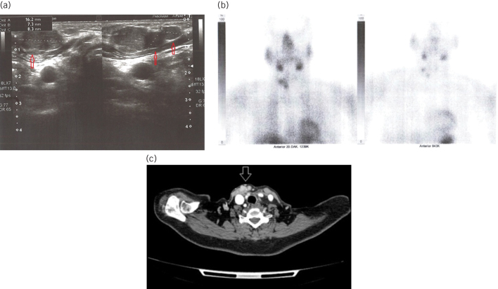
Figure 2.
a) Subcutaneous localised parathyromatosis foci (arrows) with dimensions of 16 mm, 7 mm and 8 mm, respectively, on ultrasound. b) Images of Tc99m sestamibi scintigraphy at 0th and 20th minutes. At the 20th-minute, the initial physiological involvement is seen to be disappeared. c) Contrast-enhanced cervical tomography revealed contrast enhanced multiple parathyromatosis foci (arrows) on the anterior and medial sides of the right sternocleidomastoid muscle.

