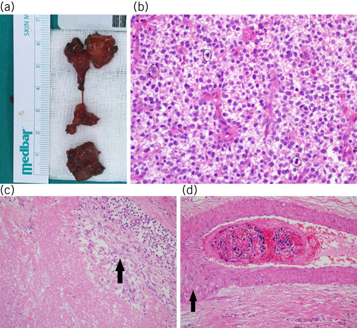
Figure 6.
a) Macroscopic appearance of the parathyromatosis foci excised from the right thyroid lodge (inferior) and left thyroid lodge (superior). b) Multiple cell mitoses (circles) were detected in parathyroid tissue (haematoxylin and eosin × 40). c) Presence of necrosis (arrow) was obtained on microscopic examination (haematoxylin and eosin × 40). d) A suspected vascular invasion image (arrow) was detected in one section (haematoxylin and eosin × 40).

