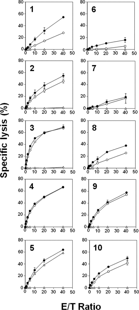
Figure 2.

Comparison of Vγ2Vδ2 T cell-mediated cytotoxicity induced by conventional N-BPs (left panels) or their fluorine-containing analogs (right panels). 786–0 renal carcinoma cells were either not treated (Δ) or pretreated with second generation N-BPs (1, 2, or 3) at 1000 μM (○) or 3000 μM (●) or their fluorine-containing analogs (6, 7 or 8) at 1000 μM (○) or 3000 μM (●) or third generation N-BPs (4 or 5) at 300 μM (○) or 1000 μM (●) or their fluorine-containing analogs (9 or 10) at 1000 μM (○) or 3000 μM (●). 786–0 renal carcinoma cells were pretreated with the N-BPs at 37oC for 2 h. The chelate-forming BM-HT prodrug reagent was then added to 25 μM and the cells incubated at 37oC for an additional 15 min. After being washed with RPMI1640 medium, the cells were cultured with Vγ2Vδ2 T cells for 40 min. Supernatants from the cultures were then harvested and mixed with Eu3+ solution. Specific lysis was determined by measuring time-resolved fluorescence using a PHERAStar multiplate reader. Data show mean ± SD and are representative of three independent experiments.
