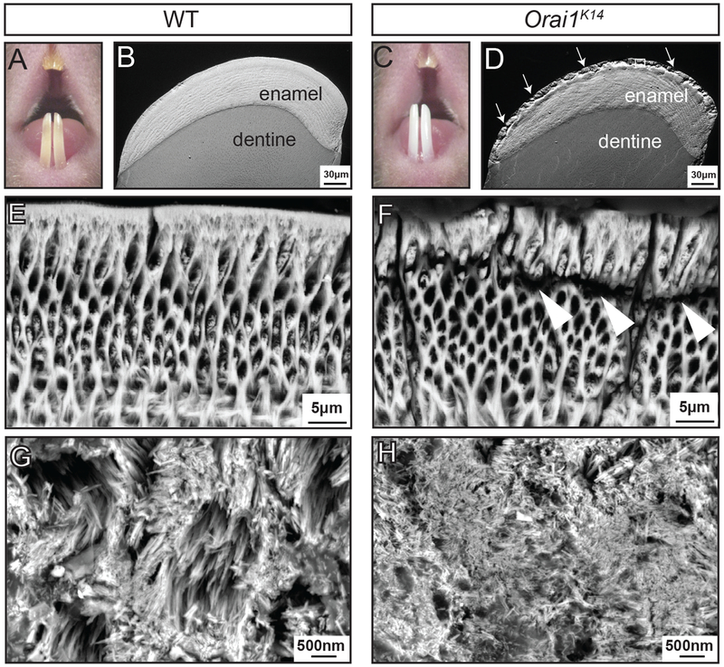
Fig. 3. Conditional deletion of Orai1 in murine ameloblast cells causes enamel defects.
(A to D) Visual examination of teeth using a stereomicroscope reveals differences in the enamel of the incisors of WT (A) and Orai1K14 (C) mice. BSE-SEM micrographs of incisor cross sections showing enamel and underlying dentine. Images were taken 1 mm from the incisor tip of WT (B) and Orai1K14 (D) mice. (E and F) High-magnification micrographs of WT (E) and Orai1K14 (F) enamel after acid etching. (G and H) FE-SEM micrographs of WT (G) and Orai1K14 (H) enamel crystals. Images shown in (A) to (H) are representative of n = 4 mice for each genotype.

