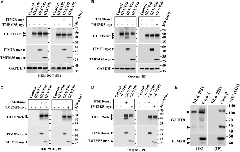
FIGURE 1.
Physical interaction between GLUT9 isoforms and ITM2B-myc or TMEM85-myc: (A) Western blot analyses of the lysates of transiently transfected HEK 293T cells, co-expressing GLUT9a/b and ITM2B/TMEM85-myc, using anti-GLUT9 antibody and anti-myc antibody respectively. (B) Western blot analyses of the lysates of microinjected oocytes co-expressing GLUT9a/b and ITM2B/TMEM85-myc. Each oocyte was microinjected with 12.5 ng of GLUT9a/b cRNA or a mixture containing 12.5 ng of GLUT9a/b cRNA and 12.5 ng of ITM2B/TMEM-myc cRNA. GAPDH protein band for each sample acts as a loading control. (C) Co-immunoprecipitation of GLUT9a/b with ITM2B/TMEM85-myc, using sepharose bead conjugated mouse anti-Myc antibody, from lysates of co-transfected HEK 293T cells co-expressing GLUT9a/b and ITM2B/TMEM85-myc. GLUT9 isoforms were detected by Western blotting using rabbit anti-GLUT9 antibody. (D) Co-immunoprecipitation of GLUT9a/b with ITM2B/TMEM85-myc, from lysates of Xenopus laevis oocytes co-expressing GLUT9a/b and ITM2B/TMEM85-myc. (E) Left panel: Western blot analyses of the lysates (60 μg total protein/lane) of HEK 293T and Caco-2 for endogenous GLUT9 and ITM2B proteins using anti-GLUT9 antibody and anti-ITM2B antibody respectively. Right panel: Endogenous GLUT9 was co-immunoprecipitated with endogenous ITM2B from the lysates of HEK 293T or Caco-2 cells using mouse anti-ITM2B antibody and sepharose (R) bead conjugated anti-mouse IgG antibody, F(ab’)2 fragment. GLUT9 was detected by immunoblotting using rabbit anti-GLUT9 antibody and ITM2B by rabbit anti-ITM2B antibody. IB, immunoblotting; IP, immunoprecipitation.

