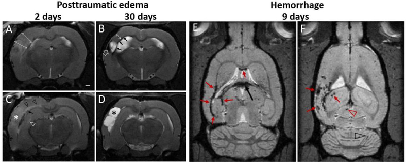
Fig. 1.
Progression of posttraumatic edema, atrophy and microbleeds. (A-D). Acute edema 2 days after LFPI (A,C) is evident in T2-wt images as hyperintensity of the contusional complex (white asterisk) and cortical swelling (double headed arrows in A). Structural MRI 2 days post-injury also shows diffuse axonal injury-related microbleeds (black arrow in C), axonal edema-associated hyperintensity of the external capsule and corpus callosum (black arrow heads), hyperintense midline of hippocampus (white arrow head) and hippocampal swelling. (B-D) In the same animal 28 days later, there is atrophy in the contusional area forming a CSF-filled cavity within the cortex (black asterisk D), cortical thickness is reduced (double arrow in B), and there is iron residues along the white matter tracts (arrows in B) as well as within the lesion (open arrow in B). Subacute hemorrhages (E-F) in T2* weighted images are shown at two horizontal levels along the white matter (red arrows in E), in choroid plexus (black arrow), as well as within the contused cortex, and in hippocampus (arrows in F), superior colliculus (red arrowhead) and cerebellum (black arrowhead). Data from EpiBioS4Rx project, courtesy of R. Immonen, University of Eastern Finland.

