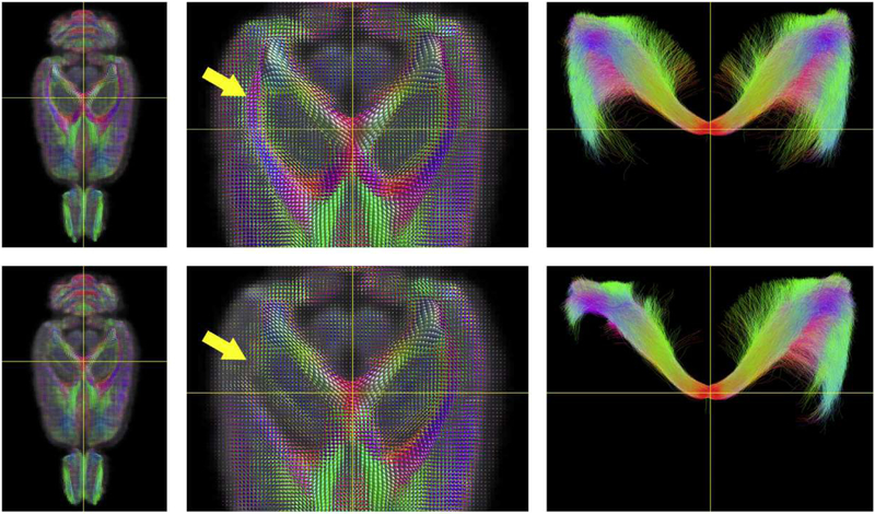
Fig. 3.
Fiber orientated distribution (FOD) and tractography template images generated for rats after sham operation (top row) and lateral fluid percussion injury (LFPI, bottom row). Damage to the white matter is reflected by reduced FOD amplitudes (yellow arrow, middle column). The FOD images were used to guide streamlines which were seeded from a spherical ROI positioned at the intersection of the two yellow lines. The resulting tractography images are shown at right and include all of the streamlines contained within the full superior/inferior slab. Following moderate LFPI, rats exhibit truncating of streamlines ipsilateral to the injury site. FODs and tractography streamlines are color-encoded according to orientation: red, medial/lateral; blue, superior/inferior; and green, anterior/posterior. Courtesy of D. Wright, Monash University.

