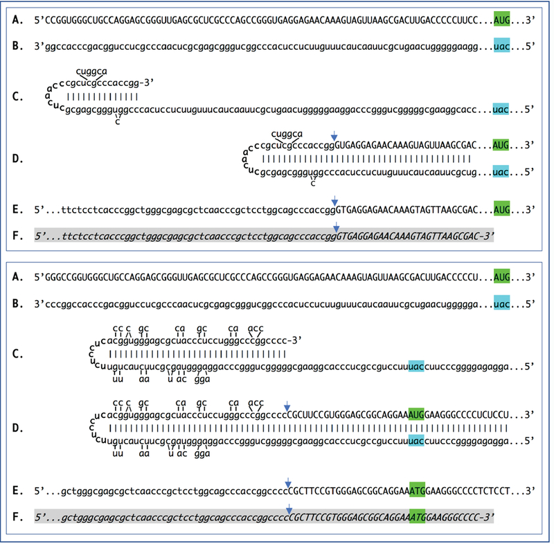
Figure 2: Sequence of a chimeric junction containing antisense and sense segments of laminin β1 mRNA and the projected pathway of its generation.

Uppercase letters – nucleotide sequence of the sense strand; lowercase letters – nucleotide sequence of the antisense strand. Highlighted in green – “AUG” translation initiation codon on the sense strand; highlighted in blue – “uac” complement of translation initiation codon on the antisense strand. In italics and highlighted in grey – detected chimeric fragments. Blue arrows: Position of antisense/sense RNA junction. The top and bottom panels depict amplification of mRNA molecules originated from different TSSs; note that self-priming positions and, consequently, chimeric junction sequences shown in two panels are different. A: 5’ terminal region of laminin pi mRNA. B: Antisense complement of the 5’ terminal region of pi laminin mRNA. C: Folding of the antisense strand into self-priming configuration. D: Extension of self-primed antisense strand into sense-oriented sequence. E: Projected chimeric junction sequence. F: Detected chimeric junction sequence. Complete sequences of the chimeric junctions shown in this Figure are provided in the main text above. Note that the priming occurs within the segment of antisense strand corresponding to the 5′UTR of mRNA, thus preserving the coding capacity of amplified mRNA.
