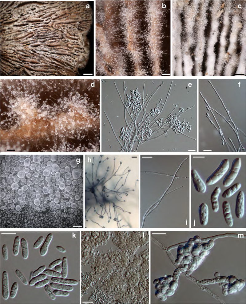
Fig. 5. Sphaerostilbella toxica.

a–d anamorph on host hymenophore; e, f, h, and i conidiogenous branches and conidia; g translucent globose mass of conidia; j, k conidia; l, m clusters of chlamydospores. a, b, d, e, and l holotype TU131905; c, f TU131904; g, h, k, m TFC202061; i, j ex–type culture TFC202258. a–f, and l on the host; h on CMD; g, i–k, m on MEA. Scale bars: a 2 mm; b, d, g 100 μm; c 300 μm; e, h 25 μm; f, i, l 20 μm; j 5 μm; k, m 10 μm
