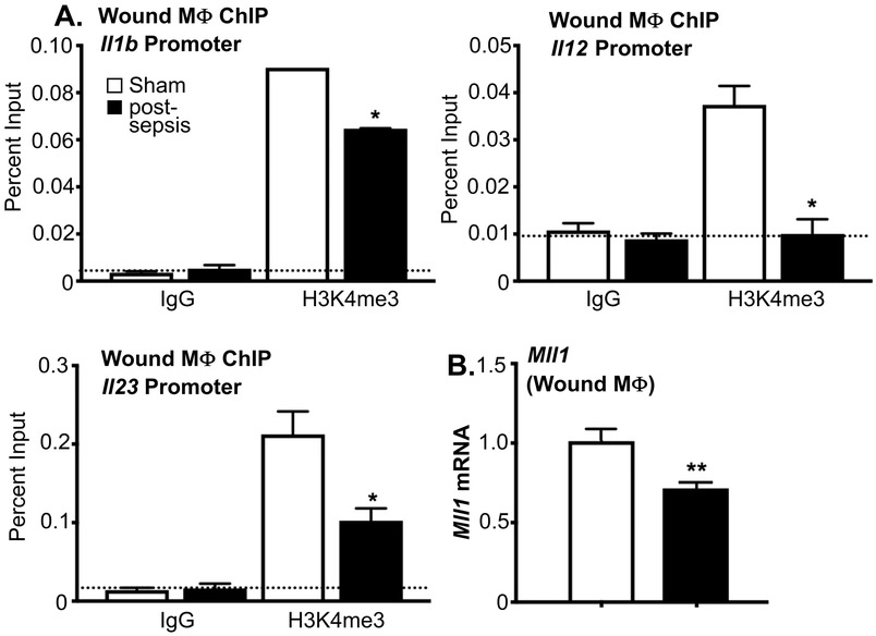
Figure 4. Wound Monocyte/Macrophages from Post-Septic Mice Demonstrate Decreased MLL1 and H3K4me3 at NFκB Binding Sites on Inflammatory Gene Promoters.
A: Wound myeloid cells were isolated at day 3 postinjury from post-sepsis and sham control mice by MACS for CD11b+[CD3-CD19-Ly6G-] cells. ChIP analysis for H3K4me3 at Il1b, Il12, and Il23 promoter was performed (n=5/group). For all ChIP experiments, isotype control antibody to IgG was run in parallel. Dotted line represents isotype control. *p<0.05 by ANOVA test with Newman-Keuls Multiple Comparison test. B: Wound myeloid cells CD11b+[CD3-CD19-Ly6G-] were isolated from post-sepsis and control mice and Mll1 expression was quantified using qPCR (n=5/group). **p<0.01 by Student t test with Welch’s correction.

