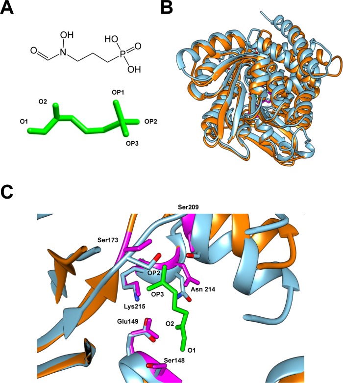
Fig 2. Comparison of the predicted structure of DxrCT and the crystal structure of DxrEC.
(A) Chemical structure (upper) and 3D structure (lower) of fosmidomycin (FSM). 3D FSM structure is in similar orientation to chemical structure, but with reactive oxygen groups labeled (“O” = oxygen; “OP” = oxygen-phosphate). (B) The superimposed view of the predicted crystal structure of DxrCT (orange) over the crystal structure of DxrEC (blue). (C) Enlarged view of the FSM binding pocket from the superimposed structures in panel B. The amino acid residues in the binding pocket of DxrEC (blue) that bind to FSM are conserved in DxrCT (labeled and highlighted in magenta). FSM is shown in green with OP2, OP3, O2, and O1 sites labeled to indicate orientation.

