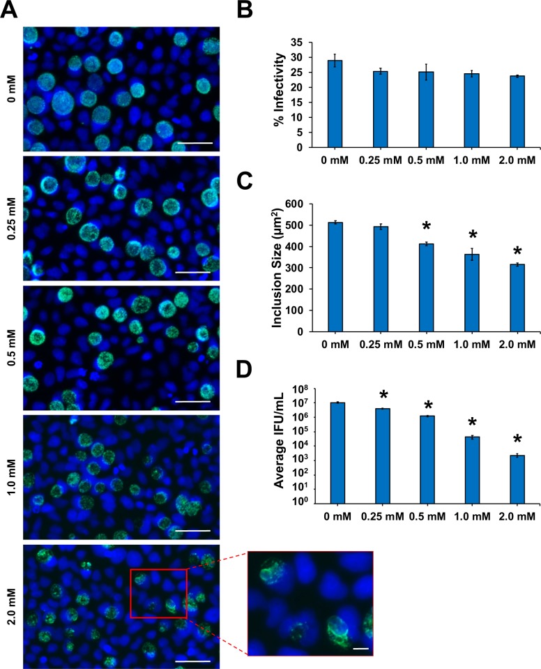
Fig 5. Chlamydial inclusion size and production of infectious EB are significantly inhibited by FSM exposure.
HeLa cells were infected with C. trachomatis at an MOI of 0.5 and harvested for analysis at 40 hpci. (A) Representative 400X magnification fluorescence microscopy images of chlamydial inclusions (green) stained with BioRad anti-chlamydial LPS Pathfinder stain and HeLa cell nuclei (blue) counter-stained with DAPI. Scale bars = 50 μm. Insert from 2 mM FSM concentration depicts zoomed-in view of ABs. Scale bars = 10 μm. (B) Percent infectivity was calculated by counting the number of inclusions/cell nuclei per field in 10 fields per coverslip in triplicate samples. (C) The area of 150 random inclusions per triplicate sample were measured using the spline contour tool in the Zen Blue Zeiss software package. (D) Production of infectious EBs was determined via chlamydial titer assays by subpassage. Significant (p ≤ 0.005) difference from untreated samples is indicated by an asterisk (*). Error bars indicate +/- SEM from triplicate samples and data are representative of three independent experiments.

