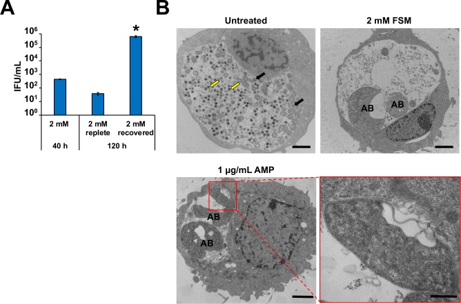
Fig 6. FSM induces persistence in Chlamydia-infected HeLa cell cultures.
At 40 hpci, cultures were washed once with complete media and either re-fed with media containing FSM (replete) or without FSM (recovered). Samples were incubated an additional 80 h and titers (A) were examined at 120 hpci. Asterisk (*) indicates significant difference from 120 h, 2 mM replete samples. Error bars indicate +/- SEM from triplicate samples and data are representative of three independent experiments. (B) Electron micrographs from untreated and 2 mM FSM-treated Chlamydia-infected HeLa cells 40 hpci. Yellow arrows and black arrows indicate normal EBs and RBs, respectively. AB indicates aberrant bodies. Scale bars = 2 μm. For AMP insert (lower right panel), scale bars = 500 μm.

