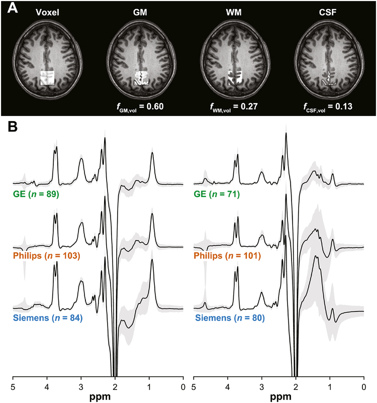
Fig. 1.
(A) Representative MRS voxel placement on a T1-weighted structural image and probabilistic partial volume voxel maps following tissue segmentation for one participant. Corresponding tissue fractions of gray matter (GM), white matter (WM) and cerebrospinal fluid (CSF) are shown. (B) Vendor-mean GABA-edited difference spectra acquired by GABA+ and MM-suppressed GABA editing. The gray patches represent ±1 standard deviation. The associated sample sizes are shown in parentheses.

