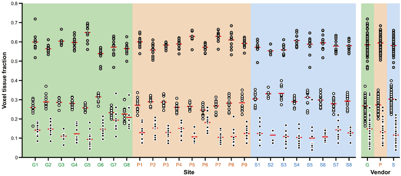
Fig. 3.
Gray matter (GM), white matter (WM) and cerebrospinal fluid (CSF) voxel tissue fractions, displayed by site and by vendor. GM = gray fill; WM = white fill; CSF = black fill. The red lines denote the mean. Sites are colored by vendor (GE sites with a green background, Philips sites with an orange background, Siemens sites with a blue background).

