Figure.

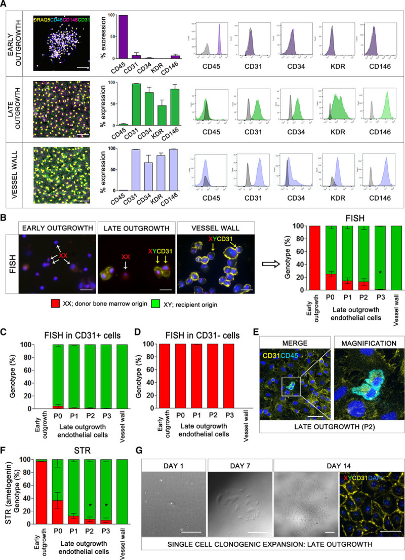
The origin of endothelial progenitor cells. A, Flow cytometric analysis of early-outgrowth cells, late-outgrowth endothelial cells, and vessel wall endothelial cells with antibodies to CD45, CD31, CD34, KDR, and CD146. Immunofluorescence staining for viable nuclei (DRAQ5, yellow), CD45 (blue), CD146 (magenta), and CD31 (green). Scale bar: 500 µm. B, Fluorescence in situ hybridization (FISH) for the X and Y chromosomes combined with CD31 staining of early-outgrowth cells (day 5), late-outgrowth endothelial cells (passage 1), and vessel wall endothelial cells (passage 1) in male patients with sex-mismatched bone marrow transplants. Representative images show Y chromosome (green), X chromosome (red), CD31 (yellow), and nuclei (DAPI, blue). Examples of CD31-negative cells with an XX genotype (white arrows) and CD31-positive cells with an XY genotype (yellow arrows) are shown. Scale bar: 20 μm. *P<0.001, one-way ANOVA. Genotype of early-outgrowth cells, late-outgrowth endothelial cells, and vessel wall endothelial cells with (C) and without (D) CD31 expression. E, Immunofluorescence for CD45 (blue) and CD31 (yellow) in late-outgrowth endothelial cells. Scale bar: 50 μm. F, Short tandem repeat analysis for the sex-specific locus, amelogenin. The proportion of cells with XX and XY genotype was calculated from polymerase chain reaction products of 104 and 110 base pairs corresponding to the X and Y chromosomes, respectively. *P<0.05, one-way ANOVA. G, Images showing expansion of a colony of late-outgrowth endothelial cells from a single endothelial progenitor cell of recipient origin (XY genotype). Y chromosome (green), X chromosome (red), CD31 (yellow), and nuclei (DAPI, blue). Scale bar: 100 μm. DAPI indicates 4′,6-diamidino-2-phenylindole; and STR, short tandem repeat.
