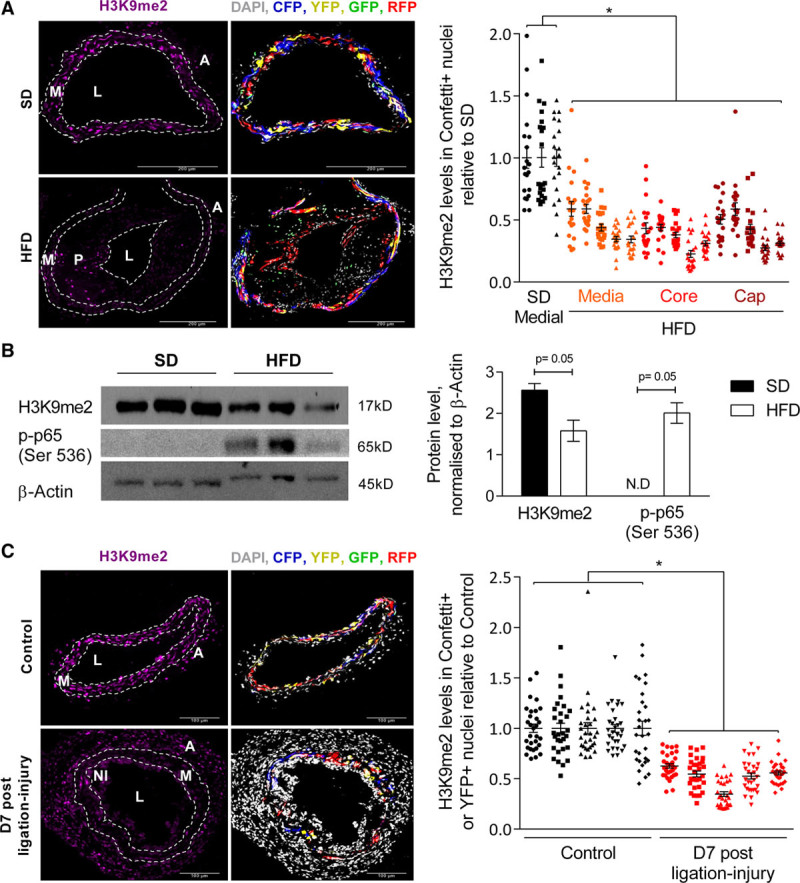
Figure 1.

Histone H3 lysine 9 dimethylation (H3K9me2) is reduced in vascular smooth muscle cells (VSMCs) in atherosclerosis and vascular remodeling. A, Representative immunofluorescence images and quantification of H3K9me2 signal intensity in nuclei of Confetti+ cells within left common carotid arteries (LCCAs) from lineage-labeled Myh11-CreERt2+/Rosa26-Confetti+/Apoe−/− males fed a standard (SD, n=3 animals) or high-fat diet (HFD, n=5 animals). H3K9me2 quantification is displayed separately for medial, plaque core, and cap VSMCs in HFD mice. B, Western blot and quantification of H3K9me2 and p-p65 (Ser 536) in aortic media from SD and HFD animals, normalized to β-Actin; n=3 animals per group. N.D., not detected. P were calculated as described in the online-only Data Supplement. C, Representative immunofluorescence images and quantification of H3K9me2 signal intensity in nuclei of Confetti+ cells within LCCAs from ligated (7 days post-ligation) relative to no surgery control Myh11-CreERt2+/Rosa26-Confetti+ mice. n=5 animals per group. A and C, Signals for H3K9me2 (magenta), Confetti reporter proteins (red, blue, yellow, green) and DAPI (4′,6-diamidino-2-phenylindole; white) are shown. The dot plots show H3K9me2 intensity in individual nuclei (of Confetti+ cells) relative to the average H3K9me2 signal intensity in control animals (SD in A, nonligated control samples in C) analyzed in the same batch (batches are indicated with symbols). Mean (line) and SEM (error bars) are indicated. *P<0.05 (linear model, see online-only Data Supplement). A indicates adventitia; L, lumen; M, media; NI, neointima; and P, plaque.
