Figure 6.

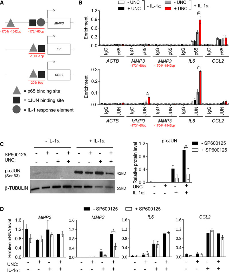
IL (interleukin)-1α-induced binding of p65 and cJUN at histone H3 lysine 9 dimethylation (H3K9me2)-target genes is enhanced by G9A/GLP (G9A-like protein) inhibition. A, Schematic indicating positions of reported p65 and cJUN binding sites and an IL-1-responsive element at the MMP3 (Borghaei et al, Clark et al, Quinones et al),41–43 IL6 (Gomard et al),44 and CCL2 (Sutcliffe et al)49 promoters. Red lines indicate chromatin immunoprecipitation (ChIP)-qPCR amplicons. B, ChIP-qPCR analysis of p65 and cJUN binding relative to input in untreated control (white bars), UNC (UNC0638; black), IL-1α (gray), and UNC+IL-1α-treated human aortic VSMCs (hVSMCs; red) showing enrichment levels at the reported binding sites within the MMP3, IL6, and CCL2 promoters and the ACTB negative control locus compared with negative control IgG (immunoglobulin). Background levels of p65 binding, indicated by a line, are based on enrichment at ACTB. Graph shows mean±SEM of 4 experiments. C, Representative western blot and densitometric analysis p-cJUN (Ser63) in untreated control hVSMCs and cells stimulated with IL-1α, without and with prior treatment with UNC and SP600125 for 48 h. Data (mean±SEM of 4 experiments) are relative to +UNC+IL-1α-SP600125 and normalized to β-TUBULIN levels. D, RT-qPCR (reverse transcription with quantitative polymerase chain reaction) analysis of MMP2, MMP3, IL6, and CCL2 in control and IL-1α-treated hVSMCs, without and with prior UNC and/or SP600125 treatment. Data (mean±SEM of 4 experiments) are relative to +UNC+IL-1α−SP600125 and normalized to the average of 2 housekeeping genes (HPRT1 and YWHAZ). Each experiment used hVSMCs derived from different individuals; *P<0.05 (Kruskal-Wallis).
