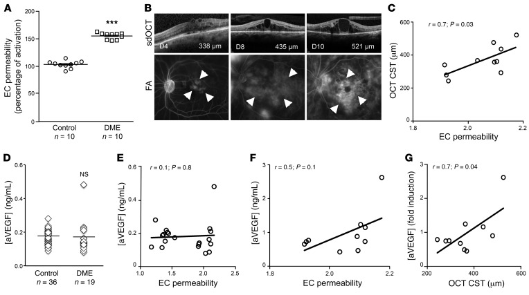
Figure 1. Vasoactive factors in the aqueous fluid from DME patients besides VEGF promote EC permeability in vitro and correlate with ME in patients.
(A) Induction of in vitro EC permeability by aqueous samples from nondiabetic (control) patients and diabetic patients with active DME (see Supplemental Table 1). DME patients show a 52% mean increase in the induction of EC permeability compared with control patients. (B) Correlation of vascular permeability with the CST on sdOCT for 3 representative DME patients (D4, D8, D10). FA, fluorescein angiogram. Arrowheads indicate leakage of fluorescein dye from retinal vasculature. (C) Correlation of the promotion of EC permeability by aqueous fluid from DME patients with their CST on sdOCT, r = 0.7; P = 0.03. (D) Levels of VEGF in aqueous samples from nondiabetic (control) patients and diabetic patients with DME who have not previously received anti-VEGF therapy or have not received anti-VEGF therapy for 12 weeks or longer in the sample eye (see Supplemental Table 2) (E) Correlation of EC permeability with levels of VEGF in aqueous samples from diabetic patient with DME. r = 0.1; P = 0.8 (E). (F and G) Correlation of EC permeability (r = 0.5; P = 0.1) (F) and CST on sdOCT (r = 0.7; P = 0.04) (G) with levels of VEGF in aqueous samples from diabetic patients with DME (see Supplemental Table 2). Two-tailed unpaired Student’s t test (A), Mann-Whitney U test (D), Pearson’s correlation (C, E, F, G). ***P < 0.001.

