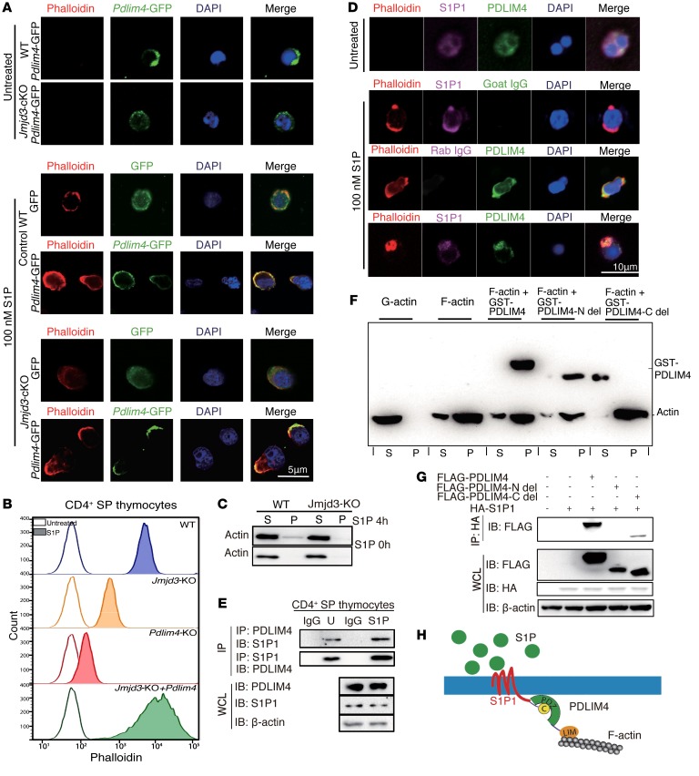
Figure 4. PDLIM4 regulates T cell migration by interaction with S1P1 and modulation of F-actin reorganization.
(A) Immunofluorescence microscopy images of WT and Jmjd3-cKO CD4+ T cells infected with lentivirus containing GFP or PDLIM4-GFP plasmids. Cells were starved 12 hours and treated with 100 nM S1P for 3 hours at 37°C. GFP or PDLIM4-GFP was detected with green fluorescence. Actin filaments were labeled with rhodamine-conjugated phalloidin (red). Nuclei were stained with DAPI (blue). (B) FACS analysis of phalloidin-labeled F-actin in untreated and S1P-treated WT, Jmjd3-cKO, Pdlim4-KO, and Pdlim4-expressing Jmjd3-cKO CD4 SP thymocytes. (C) F-actin (pellet [P]) and G-actin (supernatant [S]) from untreated and treated WT, Jmjd3-cKO CD4 SP thymocytes were detected by Western blot. (D) Immunofluorescence microscopy of untreated and S1P-treated WT CD4+ SP cells stained with FITC-conjugated Abs to detect PDLIM4, Cy5-conjugated Abs to detect S1P1, and rhodamine-conjugated phalloidin to detect actin filaments. Goat IgG and rabbit IgG were used as isotype controls. Merged images indicate colocalization of proteins. (E) Co-IP analysis of endogenous interaction of PDLIM4 with S1P1 in untreated and S1P-treated CD4 SP thymocytes. (F) Cosedimentation assay was performed using GST-PDLIM4, GST-PDLIM4 N-del, or GST-PDLIM4 C-del with F-actin, and subsequent analysis of supernatants and pellets by Western blot analysis. (G) 293T cells were cotransfected with FLAG-Pdlim4-N-del, C-del, or full-length Pdlim4 along with HA-S1p1. WCLs were immunoprecipitated with anti-HA beads and immunoblotted with anti-FLAG Ab. Three independent experiments were repeated with similar results. (H) A schematic diagram of the proposed model showing that PDLIM4 interacts with the S1P1 protein at the N-terminal PDZ domain and binds F-actin by the C-terminal LIM domain.

