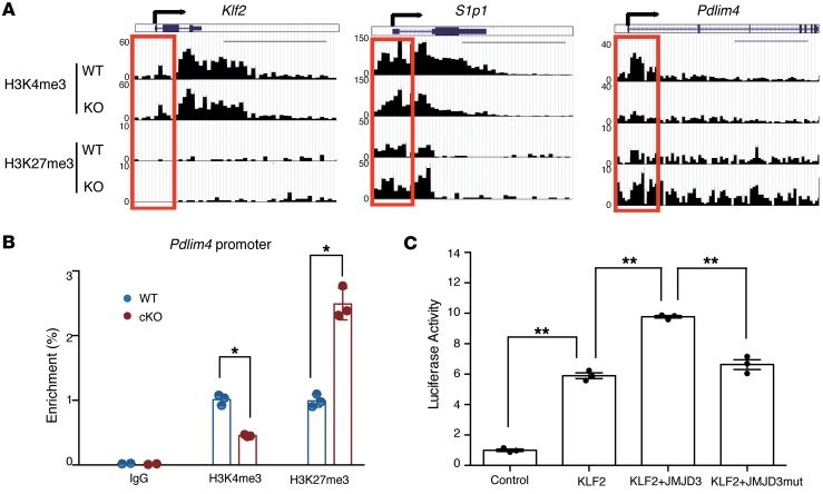
Figure 6. H3K27me3 and H3K4me3 levels in the Pdlim4 promoter in WT and Jmjd3-deficient T cells.
(A) ChIP-Seq analysis of H3K27me3 and H3K4me3 levels in the promoter and gene-body regions of Klf2, S1p1, and Pdlim4 in thymic CD4 SP T cells isolated from WT and Jmjd3-cKO mice. Red boxes indicate 2 kb regions around the TSS. Scale bars: 5 kb. (B) Validation of methylation changes on the Pdlim4 gene in WT and Jmjd3-cKO CD4 SP T cells by ChIP-qPCR. Data represent mean ± SD from 3 independent experiments. n = 3. *P < 0.05, Student’s t test. (C) Luciferase assay was performed on 293T cells cotransfected with Pdlim4 promoter–linked episomal luciferase vector and with Klf2 in the presence of WT or mutant Jmjd3 (a loss of demethylase function mutation). Data are presented as mean ± SD from 3 independent experiments. n = 3. **P < 0.01, 1-way ANOVA with Tukey’s multiple comparisons test.

