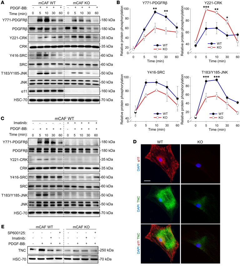
Figure 9. Integrin α11 regulates PDGFRβ downstream activation and promotes tenascin C expression in CAFs.
(A) Western blot of protein phosphorylation for PDGFRβ (Y771), CRK (Y221), SRC (Y416), and JNK (T183/S185) after 0, 5, 10, 30, and 60 minutes of PDGF-BB (10 ng/mL) stimulation in WT and KO mCAFs. (B) Quantified kinetics of PDGFRβ, CRK, SRC, and JNK protein phosphorylation from A. Data are presented as normalized ratio between phosphorylated and total proteins. n = 7 (Y771-PDGFRβ); n = 4 (Y221-CRK); n = 3 (Y416-SRC); n = 4 (T183/Y185-JNK) of independent experiments. Two-way ANOVA with Holm-Šidák multiple-comparisons test. (C) Western blot of protein phosphorylation for PDGFRβ (Y771), CRK (Y221), SRC (Y416), and JNK (T183/S185) after PDGF-BB (10 ng/mL) stimulation in WT mCAFs pretreated or not with imatinib (5 μM) for 1.5 hours. (D) Confocal immunofluorescence staining of integrin α11 (red) and tenascin C (TNC) (green) in WT and KO mCAFs. Nuclei stained with DAPI. Scale bar: 40 μm. (E) Western blot analysis of tenascin C expression before or after PDGF-BB (10 ng/mL) stimulation in WT and KO mCAFs pretreated or not with imatinib (5 μM) or SP600125 (5 μM) for 20 hours. **P < 0.01; ***P < 0.001.

