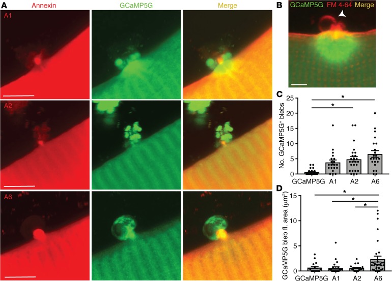
Figure 2. Annexin expression promoted release of blebs from the site if myofiber repair.
Myofibers were electroporated with the Ca2+ indicator GCaMP5G (green) with or without tdTomato-labeled annexin A1, annexin A2, or annexin A6. Ca2+ area and fluorescence were assessed after membrane damage. (A) High-magnification Z-projection images illustrate external blebs filled with the Ca2+ indicator emanating from the lesion when annexin A1, A2, or A6 was coexpressed and a corresponding reduction in Ca2+ indicator within the myofiber when compared with GCaMP5G alone (see panel B). (B) Membrane marked by FM 4-64 shows GCaMP5G-negative vesicles form in the absence of annexin overexpression. Scale bars: 5 μm. (C) Expression of annexin A6 or A2 resulted in an increased number of GCaMP5G-positive blebs. (D) Expression of annexin A6 resulted in the formation of the largest GCaMP5G-positive blebs. Data are expressed as mean ± SEM. Differences were tested by 1-way ANOVA with Tukey’s multiple-comparisons test. *P < 0.05 (n = 16 myofibers from n = 3 mice per condition).

