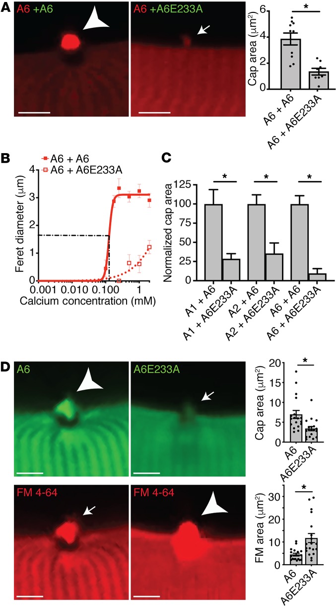
Figure 4. Annexin A6 Ca2+-binding mutant reduced annexin repair cap recruitment and decreased myofiber membrane repair capacity.
(A) Myofibers were coelectroporated with wild-type-tdTomato (red labels) and either wild-type-GFP or mutant-GFP (green labels) annexin constructs, and cap size was assessed after membrane damage; only the red channel is shown to demonstrate the effect on wild-type annexin. (B) Coexpression of mutant annexin A6E233A was sufficient to reduce wild-type annexin A6 cap assembly. Cap kinetics were plotted as cap Feret diameter over a range of Ca2+ concentrations, from 0–2 mM. (C) Coexpression of annexin A6E233A was sufficient to significantly reduce the cap area of coexpressed annexin A1, A2, and A6. *P < 0.05 for WT + WT vs. WT + mutant. (D) Myofibers were electroporated with annexin A6–GFP or mutant A6E233A–GFP. Annexin A6E233A cap area (small arrow) was significantly smaller compared with annexin A6 (large arrowhead), correlating with increased FM 4-64 fluorescence area (large arrowhead). Scale bars: 5 μm. Data are expressed as mean ± SEM. Differences were assessed by 2-tailed t test (A, C, and D). *P < 0.05 (n = 4–18 myofibers from n = 3 mice per condition).

