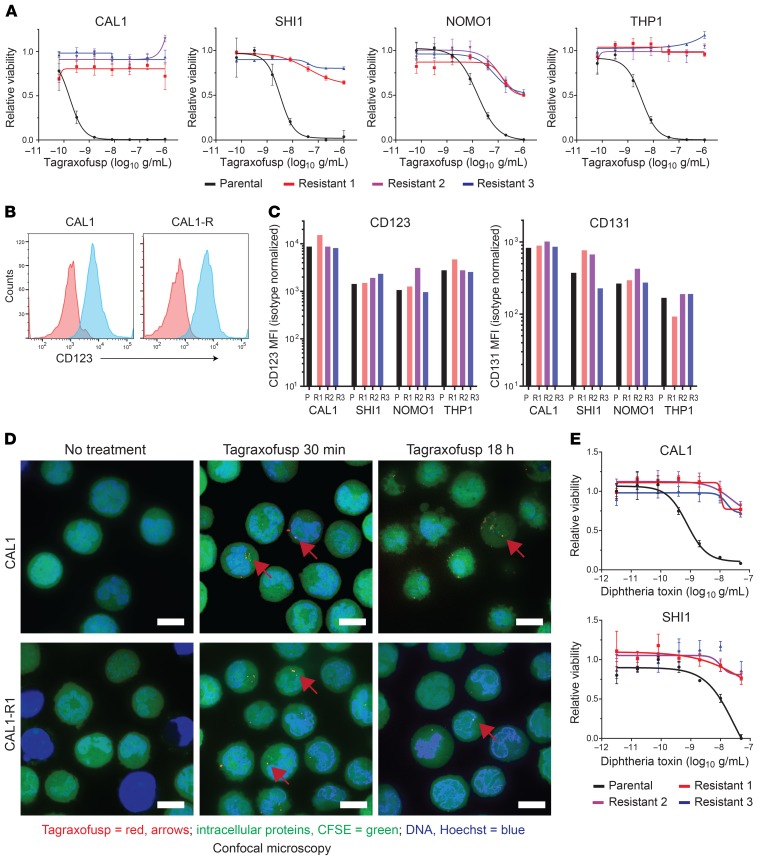
Figure 2. BPDCN and AML cells resistant to tagraxofusp maintain CD123 expression and internalization of tagraxofusp but are cross-resistant to full-length diphtheria toxin.
(A) BPDCN (CAL1) and AML (SHI1, NOMO1, and THP1) parental (black) and tagraxofusp-resistant (red, blue, purple) cultures were tested for sensitivity to 5-fold decreasing concentrations of tagraxofusp in an MTT assay. Each point was assessed in triplicate and plotted relative to cells growing in vehicle alone. (B) CD123 (blue) and isotype control (red) staining as measured by flow cytometry in parental CAL1 cells and in tagraxofusp-resistant CAL1 (CAL1-R) cells is shown. (C) MFI of CD123 and CD131 in the indicated BPDCN and AML parental (P) and tagraxofusp-resistant (R1–R3) cell lines is shown. (D) Confocal microscopy 30 minutes and 18 hours after exposure of parental or tagraxofusp-resistant CAL1 cells to APC-tagged tagraxofusp (red, representative foci highlighted by red arrows), costained with CFSE (intracellular proteins, green) and Hoechst 33342 (DNA, blue). Scale bars: 10 μm. (E) MTT assays for viability of CAL1 and SHI1 parental and 3 independent tagraxofusp-resistant subcultures after exposure to full-length diphtheria toxin (DT), plotted as in A.

