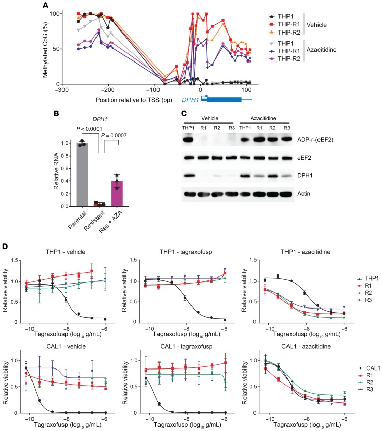
Figure 4. Tagraxofusp resistance is associated with hypermethylation of DPH1 locus CpGs, and azacitidine restores diphthamide pathway activity and tagraxofusp sensitivity.
(A) Percentage of methylated CpGs in the DPH1 locus are shown for the indicated genomic positions in parental THP1 cells and 2 independent tagraxofusp-resistant subclones, before and after 2 weeks of pulsatile treatment with noncytotoxic doses of azacitidine. (B) Quantitative RT-PCR for DPH1 expression in parental THP1 cells and a tagraxofusp-resistant subclone treated with vehicle or azacitidine (n = 3 replicates each). Dots represent relative expression, bars are ±SD. Conditions compared by 1-way ANOVA with Dunnett’s multiple-comparisons correction, adjusted P values shown. Data are representative of 2 independent resistant subclones with similar results. (C) In vitro ADP-ribosylation assay in the presence of tagraxofusp (top row) and Western blotting for eEF2, DPH1, and actin (bottom rows) are shown for parental THP1 and 3 independent tagraxofusp-resistant subclones (R1–R3) after 2 weeks of pulsatile treatment with noncytotoxic doses of azacitidine or vehicle. (D) Tagraxofusp cytotoxicity assays in parental and tagraxofusp-resistant AML (THP1) and BPDCN (CAL1) cells after 2 weeks of pulsatile treatment with noncytotoxic doses of azacitidine or vehicle, or with weekly exposure to 1 μg/mL tagraxofusp. Each point was assessed in triplicate and plotted relative to cells growing in vehicle alone.

