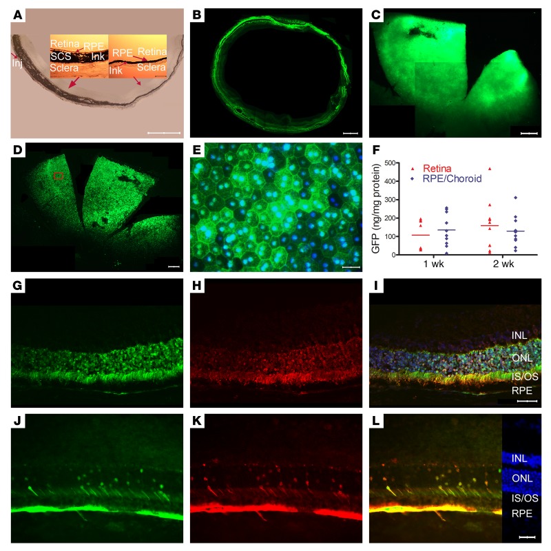
Figure 1. Widespread expression of GFP after suprachoroidal injection of AAV8.GFP in rats.
(A) Immediately after suprachoroidal injection of India ink (3 μL) in rats, frozen sections showed increased choroidal thickness on the side of injection that tapered to normal, but ink extended completely around the eye. Scale bar: 1000 μm. High magnification showed ink from the sclera to the basal surface of the RPE (insets; scale bar: 100 μm). (B) Two weeks after suprachoroidal injection of 2.85 × 10¹⁰ GCs of AAV8.GFP, 10-μm horizontal frozen sections at the equator showed GFP in the retina and RPE extending around entire circumference of the eye. Scale bar: 500 μm. (C) A retinal flat mount showed high GFP expression in about one-fifth of the retina from anterior edge posteriorly nearly to the optic nerve. Scale bar: 500 μm. (D) There was GFP throughout about one-fifth of a RPE flat mount from the anterior edge posteriorly almost to the optic nerve. Scale bar: 500 μm. (E) Higher magnification of boxed region shows strong GFP fluorescence in some RPE cells and little fluorescence in others. Scale bar: 50 μm. (F) The mean ± SEM level of GFP measured by ELISA was high in retinal or RPE/choroid homogenates at 1 and 2 weeks after suprachoroidal injection (n = 10 for each group). Two weeks after vector injection, an ocular section through the posterior part of the eye near the optic nerve shows strong fluorescence in the RPE, photoreceptor cell bodies, inner segments, and outer segments (G). The same section immunohistochemically stained with anti-GFP antibody (H) and merged with the image in G shows that the fluorescence is due to GFP (I; scale bar: 50 μm). An ocular section from the equator of the eye on the side opposite the site of injection shows strong GFP expression in RPE but in a minority of photoreceptor cell bodies and inner segments (J–L; scale bars: 50 μm). INL, inner nuclear layer; ONL, outer nuclear layer; IS, photoreceptor inner segment; OS, photoreceptor outer segment; RPE, retinal pigmented epithelium.

