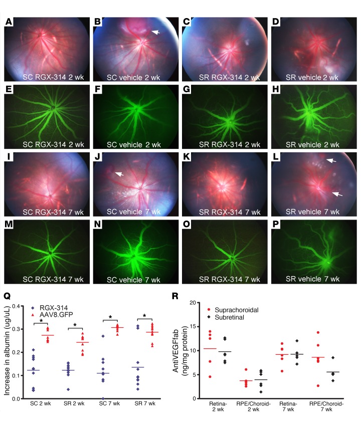
Figure 4. Suprachoroidal versus subretinal injection of RGX-314.
Rats were given suprachoroidal (SC) or subretinal (SR) injection of 1.2 × 10⁸ GCs of RGX-314 in one eye and SC or SR vehicle injection in the other eye. After 2 weeks, intravitreous VEGF (100 ng) was given and 24 hours later, fundus photographs showed normal retinas and retinal vessels in eyes given SC (A) or SR (C) injection of RGX-314, whereas SC (B) or SR (D) vehicle controls showed dilated, engorged vessels and hemorrhages (B, arrow). Fluorescein angiograms (FAs) showed normal caliber vessels with sharp margins in SC (E) or SR (G) RGX-314–injected eyes, whereas vehicle controls showed dilated vessels with blurred margins (F and H). Results were similar 7 weeks after vector injection. Twenty-four hours after VEGF (100 ng) injection, eyes that had been given SC (I and M) or SR (K and O) injection of RGX-314 showed normal retinal vessels with sharp margins in FAs, whereas vehicle controls showed dilated retinal vessels and hemorrhages (J and L, arrows), and hazy margins in FAs (N and P). (Q) Two or 7 weeks after SC or SR injection of RGX-314 or AAV8.GFP in the fellow eye, mean (± SEM) vitreous albumin measured by ELISA 24 hours after VEGF (100 ng) injection was significantly less in SC or SR RGX-314–injected eyes versus fellow eye controls (n = 10, *P < 0.03 by unpaired t test). (R) Mean (± SEM) anti–VEGF Fab in the retina and RPE/choroid were high 2 or 7 weeks after RGX-314 injection with no significant difference between the SC and SR routes of injection at each time point (n ≥ 5).

