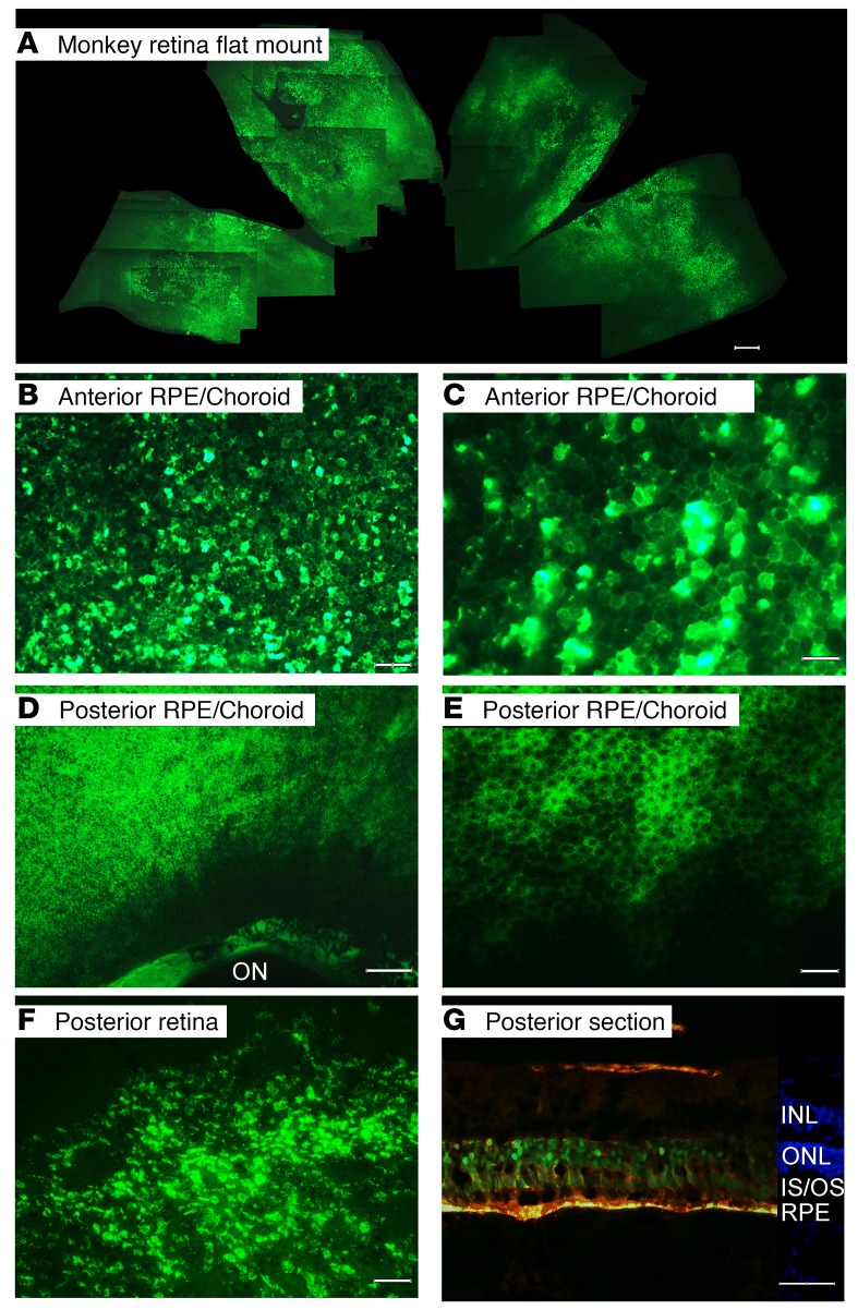
Figure 5. Widespread expression of GFP 3 weeks after suprachoroidal injection of AAV8.GFP in nonhuman primates or pigs.
Rhesus monkeys were given a suprachoroidal injection of 50 μL containing 4.75 × 10¹¹ GCs of AAV8.GFP and after 3 weeks flat mounts were examined by fluorescence microscopy. The flat mounts and sections from each of the eyes showed similar results and therefore representative images are shown. A collage was made from one eye by aligning areas of overlap. The collage shows strong expression of GFP throughout approximately one-third of the retinal flat mount (A; scale bar: 1000 μm). In a RPE flat mount, high magnification view of the mid-periphery in the quadrant of injection shows heterogeneity of GFP expression with intense fluorescence in some RPE cells and little or none in others (B; scale bar: 100 μm). A higher magnification view shows the hexagonal shape of RPE cells, some seen in negative relief (C; scale bar: 50 μm). Posteriorly there is less intense, but more uniform GFP expression in RPE cells extending almost to the border of the optic nerve (ON) which is outlined by fluorescence (D; scale bar: 250 μm). Higher magnification shows the hexagonally shaped RPE cells with GFP in the cytoplasm and the nuclei in negative relief (E; scale bar: 50 μm). Retinal flat mounts showed GFP expression in many cells of the multilayered retina extending posteriorly to the cut edge of retina where it had been severed from the optic nerve (F; scale bar: 50 μm). Two weeks after suprachoroidal injection of 50 μL containing 4.75 × 10¹¹ GCs of AAV8.GFP in a pig, merged images from ocular sections showed colocalization of fluorescence and anti-GFP staining in RPE cells and photoreceptor inner and outer segments (G; scale bar: 50 μm). There is also some GFP expression in the inner retina.

