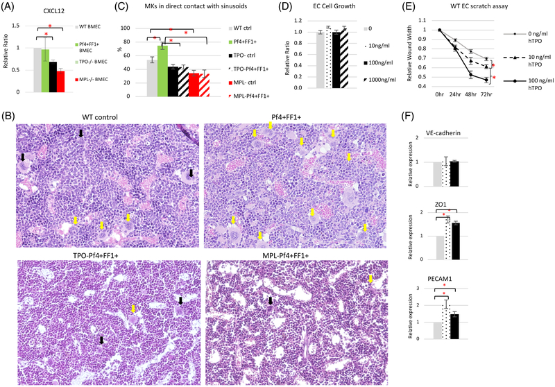
Figure 5.
TPO and MPL are important for vascular niche function. (A) The expression levels of CXCL12 in marrow ECs isolated from WT ctrl, Pf4+FF1+, TPO−/−, and MPL−/− mice. Gene expression is shown as the relative fold-change compared to WT ctrl marrow EC expression which was set as ‘1’. (B) Representative images of H&E sections of femoral marrow of Pf4-cre WT ctrl mice, Pf4+FF1+ mice, TPO−Pf4+FF1+ mice and MPL−Pf4+FF1+ mice. Yellow arrows indicate examples of MKs physically associated with marrow sinusoids. Black arrows indicate examples of MKs not in direct contact with marrow sinusoids. Magnification: x20 (C) Quantification of MKs physically associated with marrow sinusoids in WT ctrl mice (n = 3), Pf4+FF1+ mice (n = 3), TPO− ctrl mice (n = 2), TPO−Pf4+FF1+ mice (n = 2), MPL− ctrl mice (n = 2), and MPL−Pf4+FF1+ mice (n = 2). About 100 marrow MKs from 9–18 non-overlapping fields were studied. (D) TPO did not affect primary murine spleen EC cell growth in vitro. (E) TPO stimulated primary murine spleen EC cell migration in vitro. A lesion was produced across the primary murine spleen EC monolayer in the presence of different TPO concentrations. The distances from one side of the scratch wound to the other side were measured using ImageJ software (National Institute of Health, Bethesda, MD, USA) at six different locations for each culture condition. The distance of wound closure at time 24, 48, and 72 h was compared with the distance at time 0 h which was set as 1. The results were expressed as the mean ±s.e.m. (n = 6). Data are from one of two independent experiments that gave similar results. (F) The expression levels of VE-cadherin, ZO1, and PECAM1 in WT murine lung EC treated with different TPO concentrations. Data are presented as mean ±SEM. *P < 0.05.

