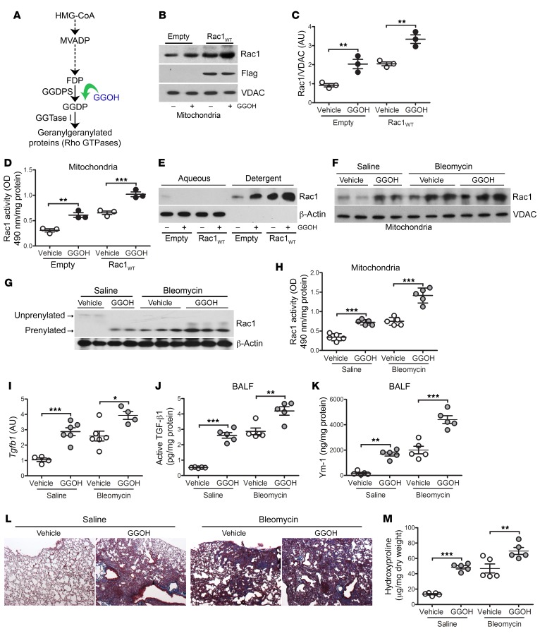
Figure 2. Increasing Rac1 activity by augmentation of isoprenylation promotes lung fibrosis.
(A) Schematic diagram of the mevalonate pathway. HMG-CoA, 3-hydroxy-3-methylglutaryl coenzyme A; MVADP, mevalonate 5-diphosphate. (B) Immunoblot analysis and (C) quantification of isolated mitochondria from THP-1 cells expressing empty control or Rac1WT and treated with vehicle or GGOH (50 μM) (n = 3). (D) Mitochondrial Rac1 activity in transfected MH-S cells treated with vehicle or GGOH (n = 3). (E) Immunoblot analysis of transfected macrophages expressing empty control or Rac1WT and treated with vehicle or GGOH. Cells were separated into aqueous (unprenylated) or detergent (prenylated) fractions. Ten days after exposure of WT mice to saline or bleomycin, pumps containing vehicle or GGOH were implanted s.c., and the mice were sacrificed 11 days later. (F) Mitochondrial Rac1 immunoblot analysis of isolated MDMs. (G) Isoprenylation status of Rac1 in isolated MDMs. (H) Mitochondrial Rac1 activity (n = 5/group). (I) Tgfb1 mRNA expression (saline, vehicle n = 4; saline, GGOH n = 6; bleomycin, vehicle n = 6; bleomycin, GGOH n = 4). (J) Active TGF-β1 and (K) Ym-1 expression in BALF (n = 5/group). (L) Representative lung histology images with Masson’s trichrome staining (n = 5/group). Original magnification, ×2.5. (M) Hydroxyproline content (n = 5/group). Values indicate the mean ± SEM. *P < 0.05, **P < 0.001, and ***P < 0.0001, by 1-way ANOVA followed by Tukey’s multiple comparisons test.

