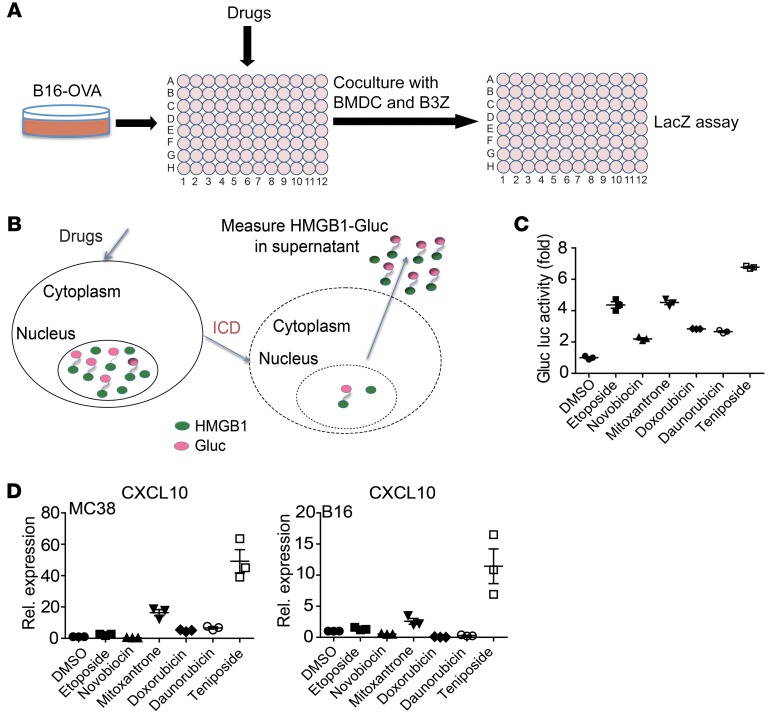
Figure 1. T cell–based drug screening identified ICD inducers.
(A) Outline of drug-screening protocol. B16-OVA tumor cells were seeded on 96-well plates and treated with drugs for 16 hours, then cocultured with BMDC and B3Z cells for 24 hours. LacZ reporter activity was measured as a surrogate marker for T cell activation. (B) Illustration of the principle of the HMGB1-Gluc reporter system. Once drugs or inhibitors induce tumor cell ICD, HMGB1-Gluc is released from the nucleus into the supernatant, and supernatant luciferase activity is detected. (C) MC38 (HMGB1-Gluc) cells were treated with different Top inhibitors or DMSO for 20 hours; then HMGB1-Gluc luciferase (Gluc luc) activity was measured. (D) MC38 and B16 cells were treated as in C, and then the mRNA expression level of CXCL10 was measured by qPCR. Rel., relative. Data in C and D are shown as mean ± SD of 3 independent experiments.

