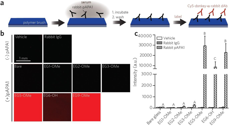
Figure 2 |.
Screening POEGMA bottlebrush surfaces for immune reactivity toward a polyclonal APA (pAPA1). a, Schematic of pAPA1 fluoroimmunoassay. Surfaces were incubated with a solution of rabbit-derived pAPA1 spiked into undiluted calf serum, rinsed, and then labeled with Cy5-donkey-α-rabbit dAbs, and then imaged with a fluorescence scanner. b, c, Representative Cy5 channel fluorescence images (b) and quantitation of mean fluorescence intensities (c). Results in (c) are plotted as mean ± 95% CI for n ≥ 4 replicates. Vehicle-only and rabbit-IgG (not specifically reactive to PEG) controls are also included to show baseline values. Incubating surfaces with pAPA1 leads to significant fluorescence, indicating APA binding, from EG5-OMe, EG6-OH, and EG9-OMe surfaces, but not from bare, EG1-OMe, EG2-OM2, or EG3-OMe surfaces. Bars marked with different letters indicate significant differences within the pAPA1-treated groups by multiple comparison testing in one-way ANOVA (Tukey post hoc test, p ≤ 0.05).

