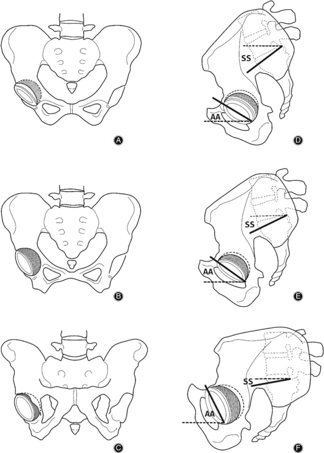
Figure 4.

Coronal and sagittal views of the pelvis with the patient supine, standing, and sitting. AA, acetabular anteversion; ST, sacral slope. (A–C) Coronal view of the pelvis, showing coronal pelvic tilt and acetabular anteversion. From supine to standing, and to siting, pelvis is gradually tilted backwards and acetabular anteversion is gradually increasing. (D–F) Sagittal view of the pelvis, showing sagittal pelvic tilt and acetabular anteversion. From supine to standing, and to siting, pelvis is gradually tilted backwards and acetabular anteversion is also gradually increasing.
