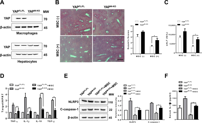
Figure 3. Myeloid YAP deficiency in MSC-treated livers aggravates IR-induced hepatocellular damage and promotes NLRP3 inflammasome-driven inflammatory response.

The YAPFL/FL and YAPM-KO mice were subjected to 90min of partial liver warm ischemia, followed by 6h of reperfusion. Some animals were injected via tail vein with MSCs (1×10⁶) 24h prior to ischemia. (A) The YAP expression was detected in hepatocytes and liver macrophages (Kupffer cells) by Western blot assay. Representative of three experiments. (B) Representative histological staining (H&E) of ischemic liver tissue (n=4–6 mice/group) and Suzuki’s histological score. Scale bars, 100μm. (C) Hepatocellular function was evaluated by sALT levels (IU/L) (n=4–6 samples/group). (D) qRT-PCR-assisted detection of TNF-α, IL-1β, IL-10, and TGF-β in ischemic livers (n=3–4 samples/group). Data were normalized to HPRT gene expression. (E) Immunoblot-assisted analysis and relative density ratio of NLRP3 and cleaved caspase-1 in ischemic livers. Representative of three experiments. (F) ELISA analysis of IL-1β levels in animal serum (n=3–4 samples/group). All data represent the mean±SD. *p<0.05, **p<0.01.
