
Figure 7. YAP is crucial to mediate β-catenin activity and reprograms NLRP3-dependent macrophage polarization in MSC-mediated immune regulation.
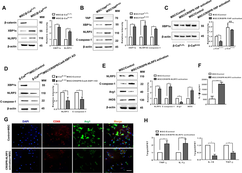
(A) (B) BMMs were isolated from β-cateninFL/FL, β-cateninM-KO, YAPFL/FL, YAPM-KO mice and then co-cultured with MSCs followed by LPS stimulation (n=3–4 samples/group). Immunoblot-assisted analysis and relative density ratio of macrophage β-catenin, YAP, XBP1s, and NLRP3, and cleaved caspase-1. Representative of three experiments. (C) BMMs were isolated from β-cateninFL/FL and β-cateninM-KO mice and transfected with CRISPR-mediated YAP activation or control vector, and then co-cultured with MSCs followed by LPS stimulation. Immunoblot-assisted analysis and relative density ratio of macrophage XBP1s. Representative of three experiments. (D) BMMs were isolated from β-cateninM-KO mice and transfected with CRISPR/Cas9-mediated XBP1 KO or control vector and then co-cultured with MSCs followed by LPS stimulation. Immunoblot-assisted analysis and relative density ratio of macrophage XBP1s, NLRP3, and cleaved caspase-1. Representative of three experiments. Fig. 7E-H, BMMs were isolated from β-cateninFL/FL mice and transfected with CRISPR-mediated NLRP3 activation or control vector were co-cultured with MSCs followed by LPS stimulation. (E) Immunoblot-assisted analysis and relative density ratio of macrophage NLRP3, cleaved caspase-1, Arg1, and iNOS. Representative of three experiments. (F) ELISA analysis of IL-1β levels in animal serum (n=3–4 samples/group). (G) Representative immunofluorescence staining for the macrophage marker CD68 (red) and arginase-1 (Arg1, green) co-localization in BMMs. DAPI was used to visualize nuclei (blue). Scale bars, 20μm. (H) qRT-PCR-assisted detection of TNF-α, IL-1β, IL-10, and TGF-β in macrophages (n=3–4 samples/group). Data were normalized to HPRT gene expression. All data represent the mean±SD. *p<0.05, **p<0.01.