
Fig. 2.
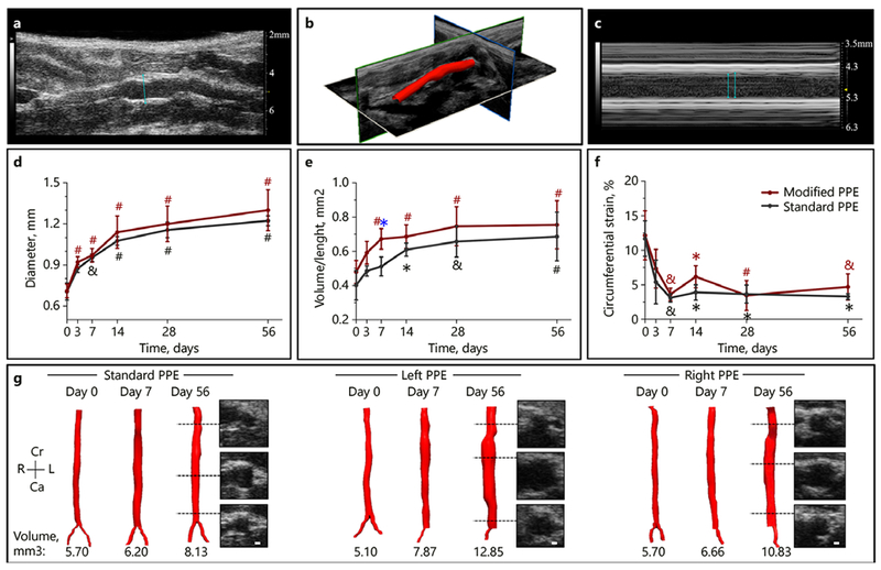
Summary of structural changes in murine abdominal aortic aneurysm development among standard, left, and right PPE groups. Long-axis B-mode was used to quantify aortic diameter (a), 3D ultrasound for aortic segmentation and volume characterization (b), and M-mode to quantify systolic and diastolic diameters (c) that were used to assess changes in circumferential cyclic strain. The aortic diameter continues to increase up to day 56 (d) with the modified PPE group having a rapid increase in volume/length up to day 7 (e). We also observed a sharp decrease in circumferential cyclic strain between baseline and day 7 post-procedure time points (f). Representative short-axis ultrasound, 3D segmentation, and volume showed slightly larger aneurysm in the modified PPE group compared to the standard PPE group (g). Statistical significance compared with day 0 and defined at * p < 0.05, & p < 0.01, and # p < 0.001. The blue asterisk (e) represents statistical significance between the standard and modified PPE groups.