
Fig. 5.
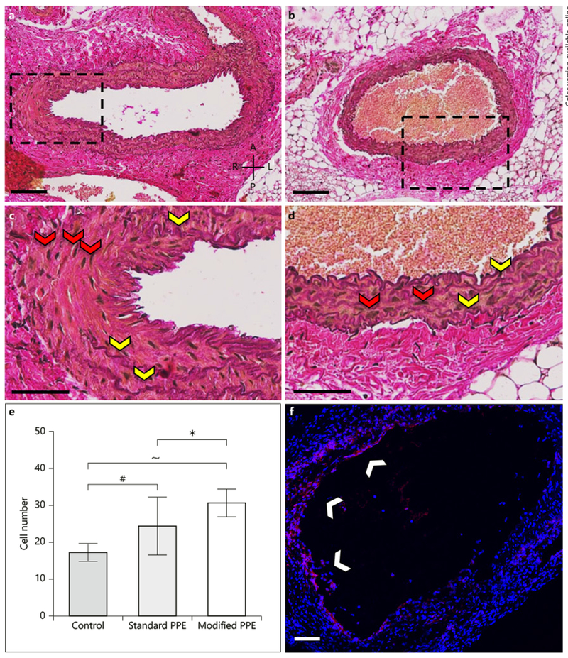
Histological and immunohistochemistry analysis of PPE-infused aortic tissue consisting of EvG-stained standard (a, c) and modified (b, d) aortae, as well as Ki67-stained modified (f) infrarenal aorta. Histology shows diffuse elastin breakage throughout the vessels (yellow arrows), thus confirming aneurysm induction. Ki67 staining shows cell proliferation in the standard and modified PPE animals (white arrows). Further, cell number analysis showed a significant increase in cell nuclei (red arrows) between the modified and standard PPE-infused aortae (h). A, anterior; P, posterior; L, left; R, right. Scale bars = 100 μm (a, c) and 50 μm (b, d). Statistical significance is defined at * p < 0.05, # p < 0.001, ~ p < 0.0001.