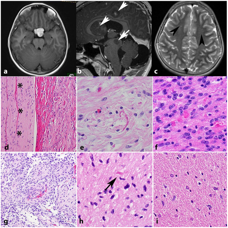
Figure 3. Low grade astrocytomas in NF1.
The optic pathways, including the optic chiasm are favored sites for low grade astrocytomas in NF1. Axial T1 weighted MR image in a 41-year-old woman with NF1 demonstrates a bulky mass involving the optic chiasm (a) that histologically proved to be a pilocytic astrocytoma. Multicentricity (arrows) of pilocytic astrocytomas is a feature of NF1 (arrows) (b). “Unidentified bright objects” represent hyperintensities on MRI that do not grow and frequently regress over time (arrowheads). They are frequent in individuals with NF1 and should not be misinterpreted as tumors (Axial T2-weighted image) (c). Pilocytic astrocytomas involving the optic nerve (asterisks) (optic nerve glioma) frequently extend into the subarachnoid space (d). Most gliomas in individuals with NF1 are pilocytic astrocytomas, characterized by frequent Rosenthal fibers (e) and eosinophilic granular bodies (f). The pilomyxoid variant of pilocytic astrocytoma also may develop in individuals with NF1 (g). Low grade astrocytomas that are difficult to classify, usually having infiltrative features but with occasional piloid features such as rare Rosenthal fibers (arrow) are also relatively frequent (h). Diffuse astrocytomas also occur in individuals with NF1 and resemble sporadic diffuse astrocytomas, particularly single cell infiltration and lack of Rosenthal fibers(i).

