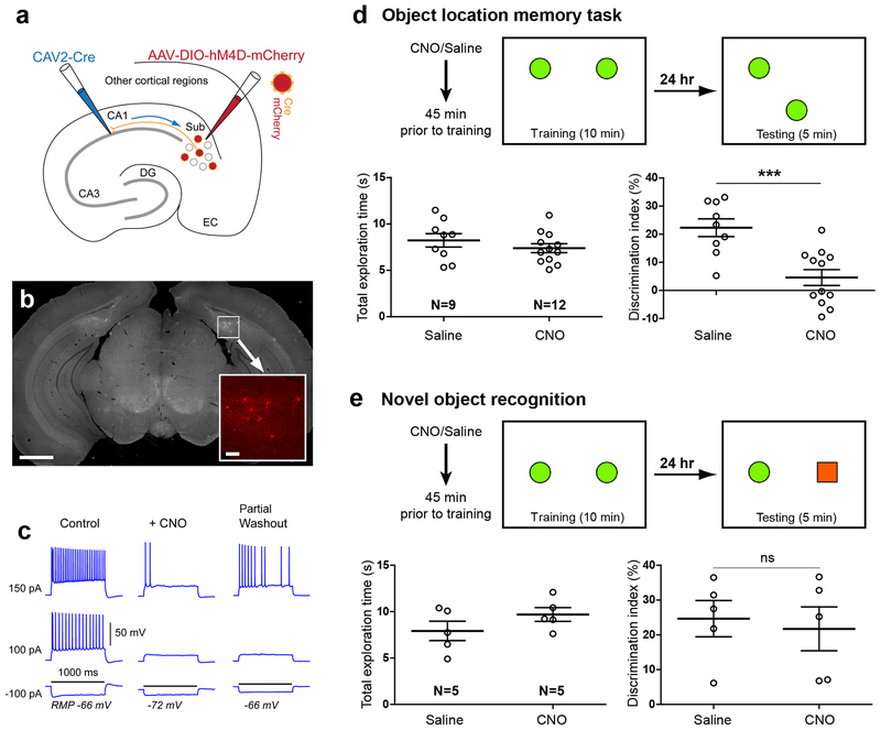
Fig. 3. Genetically targeted inactivation and activation of CA1-projecting excitatory SUB neurons modulates object-location memory.
a, Schematic illustration of genetic inactivation of CA1-projecting SUB excitatory neurons using dual CAV2-Cre injection in CA1 and AAV2-DIO-hM4D-mCherry injection in the SUB. b, Histological analysis in coronal brain sections verifies bilateral, spatially restricted hM4D-mCherry expression in SUB. Scale bar = 1 mm. The bottom right insert shows a higher magnification view of the white square region in b with hM4D-mCherry expressing, CA1-projecting SUB neurons in red. The insert scale bar = 100 μm. The viral injection experiment was independently repeated in 21 mice, each with similar results. c, Example electrophysiological data demonstrating in vitro validation of DREADDs-mediated SUB neuronal inactivation. The 5 μM CNO application hyperpolarized the cell’s resting membrane potential (RMP) and suppressed action potential firing by intrasomatic current injections. The black horizontal line indicates 1000 ms of the current injection duration with different strengths (i.e., −100 pA, 100 pA and 150 pA). The 5 μM concentration matched the CNO dosage of 1.5 mg/kg used for in vivo DREADDs experiments. Given hM4D is a G-protein coupled receptor, CNO effects can only be partially reversed with washout of 30–45 minutes, which is consistent with published results. The experiment was independently repeated in 10 cells from 4 mice, each with similar results. See more validation data in Supplementary Fig. 4a–c. d. Scheme for experimental design and results of location-dependent object recognition task following CNO-activated inhibition of CA1-projecting SUB excitatory neurons during the training phase. The box represents the open field arena, and the green filled circles indicate the training (left) and test (right) object locations. Before the experiment, mice were handled and habituated to the context in the absence of objects. Mice received a single dose i.p. injection of control saline or experimental CNO treatment (1.4mg/kg) 45 min prior to the training. Left graph: total exploration time of the animals during the testing session. Right graph: discrimination index for the testing session 24 hours after training. CNO treated mice show no preference for the moved object in contrast to saline treated controls. Data are presented as mean ± SE. *** p =0.0005 (two-tailed t-test). e, Test of novel object recognition. In training sessions, two identical objects (green filled circles) are placed in the arena, while for testing, distinct objects (green filled circle and red filled square) are placed. Mice with inhibition of CA1-projecting SUB neurons show equal preference for the novel object, similar to the saline treated controls. Data are presented as mean ± SE. n.s., not significant p = 0.73 (two-tailed t-test). For more data information, see Supplementary Table 2a.

