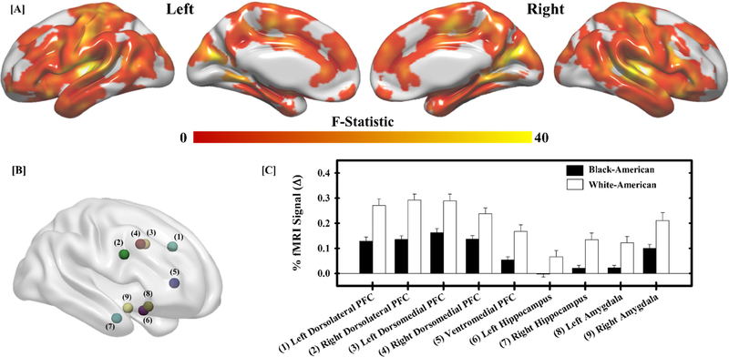
Figure 2.
Racial differences in the neural response to threat. A significant main effect of race (White-American versus Black-American) was observed across the brain (a). Follow-up analyses were completed on significant peaks of activation in regions important for threat-related emotional function (b). Within these regions, White-American participants (white bars) exhibited a greater fMRI signal response (% change) to threat than Black-American participants (black bars) (c). Bars reflect the mean fMRI signal response for each group and error bars reflect the standard error of the mean. Numbers next to region labels in (c) correspond to numbered regions in (b).

