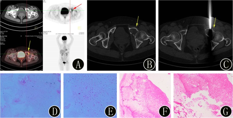
Fig. 4.
A 46-year-old woman with suspected primary bone malignancies of the pelvis. a 18F-FDG PET/CT imaging shows uptake (SUVmax 5.1) in a left pubic lesion (yellow arrow). The axial PET image also confirms the presence of the bone lesion (red arrow). b-c The intraprocedural axial noncontrast CT image (using bone windows) shows the biopsy needle targeted within the lesion (yellow arrow). The histopathologic biopsy results (d: hematoxylin and eosin, original magnification 40×, e: hematoxylin and eosin, original magnification 100×) confirmed the bone lesion as a highly differentiated chondrosarcoma. Finally, the surgical histopathology results (f: hematoxylin and eosin, original magnification 40×, g: hematoxylin and eosin, original magnification 100×) of the bone lesion confirmed the diagnosis of a highly differentiated chondrosarcoma (WHO grade I)

