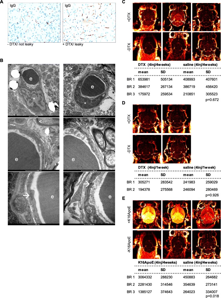
Fig. 3.
Permeability analyses of DTX-treated mice in-vivo do not show signs of increased leakage. a Exemplary IHC-stainings of murine IgG using brain sections of DTX-treated vs. control mice (original magnification 20x). b Representative images of ultrastructural NVU-imaging: ECs (indicated by *); TJs (indicated by ➔); perivascular axons (indicated by <=; mitochondria (indicated by >); basement membrane (indicated by .--.); erythrocyte (indicated by e). Images taken using Tecnai Spirit BioTWIN FEI EM at 120 kV, with 4 K CCD camera. c-e DCE-MR imaging heatmaps of treated vs. untreated mice for in-vivo permeability analysis. Statistical analysis was conducted using student’s t-test, subset analysis for three different groups as depicted in the methods section

