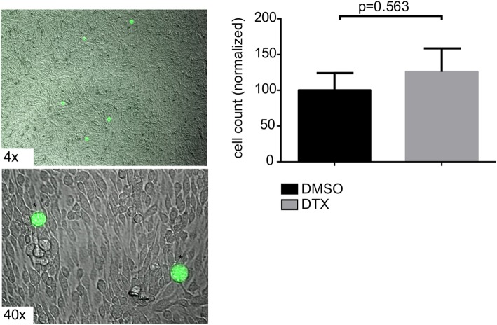
Fig. 4.
TCs do not show increased adhesion on EC monolayer upon DTX treatment. Representative images of the adhesion assay showing GFP-labeled (*) MDA-MB-231-BR-GFP-TCs on top of ECs monolayer. Phase-contrast microscope with IF-imaging, original magnification 4x, 40x. Unpaired t-test of treated (N = 3) vs. untreated (N = 3) bEnd5 cells monolayer, with TCs plated on top. Statistical analysis was done using GraphPad Prism software

