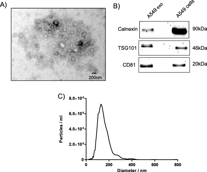
Fig. 1.
Identification and characterization of A549 cell-derived exosomes. Exosomes were isolated using differential centrifugation. a The electron micrographs of the exosomes revealed rounded structures with a size of approximately 30–150 nm. The scale bar represents 200 nm. b Western blot analysis of exosomes derived from the supernatant of A549 cells shows the presence of the common exosomes proteins CD81, calnexin and TSG101. Cells were used as a control. c The sizes of exosomes derived from the supernatant of A549 cells were analyzed using nanoparticle tracking analysis

