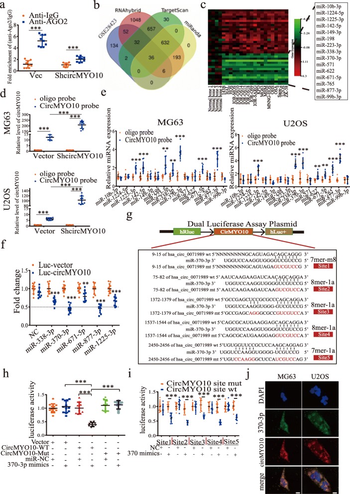
Fig. 3.
CircMYO10 acts as a sponge of miR-370-3p in osteosarcoma cells. a Ago2 RNA immunoprecipitation (RIP) assay for circMYO10 levels in MG63 cells stably expressing shcircMYO10. Data represents the mean ± SD (n = 9). b Using data from the GEO dataset GSE28423, differentially expressed miRNAs with |fold change| > 1 and p value < 0.05 were compared to the miRNAs common to the prediction of RNAhybrid, miRanda, and TargetScan that may bind to circMYO10. The Venn diagram shows the number of overlapping miRNAs. c The heat map for 36 differentially expressed miRNAs that may bind to circMYO10. d Lysates prepared from MG63 and U2OS cells stably transfected with circMYO10 or vector were subjected to RNA pull-down assays and were tested by qRT-PCR. Relative levels of circMYO10 pulled down by the circMYO10 probe were normalized to the level of circMYO10 pulled down by an oligo probe. Data represents the mean ± SD (n = 9). e The relative level of 15 miRNA candidates in the MG63 and U2OS lysates were detected by qRT-PCR. Data represents the mean ± SD (n = 9). f, h, i HEK-293 T cells were transfected with the indicated miRNA mimics and luciferase reporter plasmids. Forty-eight hours later, 293 T cells were lysed and the extracts were subjected to a dual-luciferase reporter assay. Data represent the mean ± SD (n = 9) for three independent experiments. f Luciferase activity of the Luc-vector or Luc-circMYO10 in 293 T cells co-transfected with indicated 5 miRNA mimics. g CircMYO10 was predicted to contain 5 putative sites for miR-370-3p. h 293 T cells were transfected with miR-370-3p mimics and Luciferase reporter plasmids containing either the wild type circMYO10 3′ UTR or a mutant circMYO10 3′ UTR where 5 binding sites for miR-370-3p were all mutated. i Each binding site, which was wild type or mutated, was cloned into luciferase reporter plasmids one by one. MiR-370-3p mimics were transfected into 293 T cells together with one of ten luciferase reporter plasmids separately. j FISH showed high co-localization of Cy3 labeled-circMYO10 and miR-370-3p labeled with Alexa Fluor 488 in MG63 and U2OS cells. Scale bars = 20 μm. Three independent assays were performed in the above assays. a, d-f, h-i * P < 0.05, ** P < 0.01, *** P < 0.001 (Student’s t-test)

