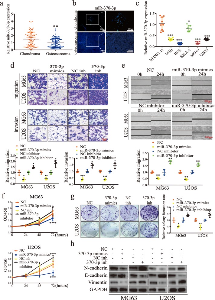
Fig. 4.
MiR-370-3p inhibits osteosarcomas cells proliferation and EMT. a The expression of miR-370-3p in ten paired chondroma (n = 10) and osteosarcoma tissues (n = 10) was measured by qRT-PCR and FISH assays. a Data represents the mean ± SD (n = 90 per group). b FISH assays showed miR-370-3p expression is lower in human OS tissue than in chondroma tissue. Representative images are shown. Scale bars = 100 μm. c The expression of miR-370-3p in hFOB1.19, 143B, U2OS, HOS, MG63, and SJSA-1 was measured by qRT-PCR. Data represents the mean ± SD (n = 9). d The effect of miR-370-3p on migration and invasion was measured in Transwell migration and invasion assays. Data represents the mean ± SD (n = 3). Scale bars =50 μm. e The effect of miR-370-3p overexpression and inhibition on migration was measured by wound healing assays. Data represents the mean ± SD (n = 3). Scale bars = 200 μm. f Proliferation of cells transfected with miR-370-3p mimics or miR-370-3p inhibitor was measured by CCK-8 assay in MG63 and U2OS cells. Data represents the mean ± SD (n = 18). g Downregulation of miR-370-3p stimulates colony formation and overexpression of miR-370-3p suppresses colony formation in MG63 and U2OS cells. Data represents the mean ± SD (n = 3). f The effect of miR-370-3p on the expression of CyclinD1, E-cadherin, N-cadherin, and vimentin was detected by western blot analysis. Three independent assays were performed in the above assays. a, c, d-f * P < 0.05, ** P < 0.01, *** P < 0.001 (Student’s t-test)

