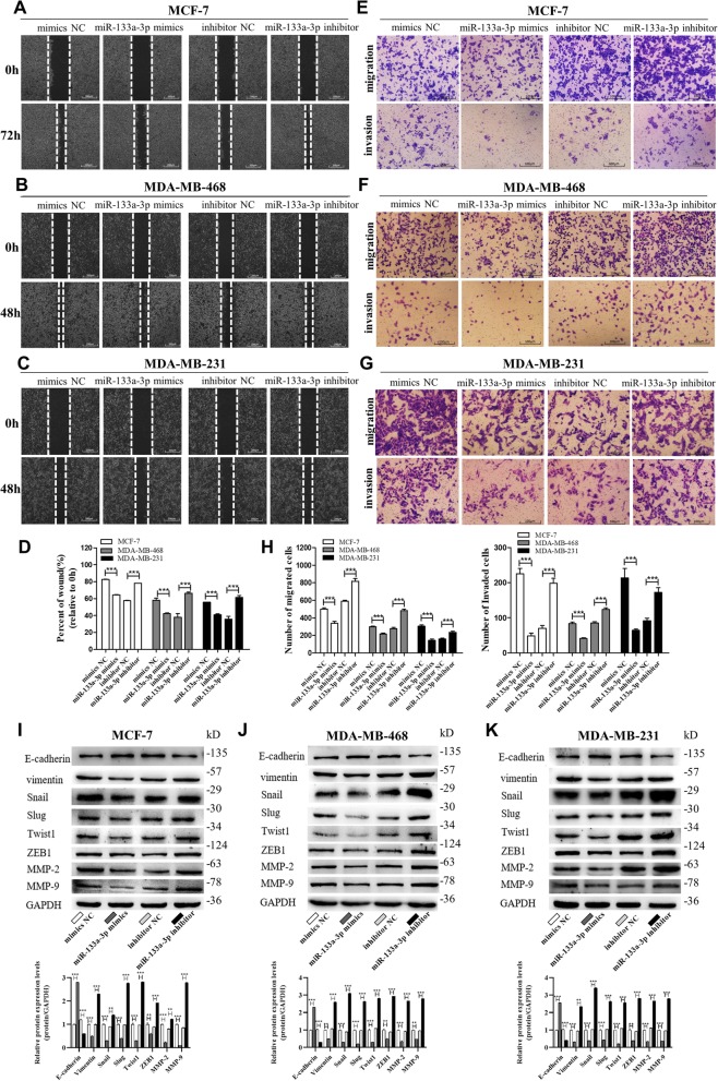
Fig. 2.
miR-133a-3p promotes breast cancer cells migration and invasion in vitro. a-d Migration of MCF-7 cells (a), MDA-MB-468 cells (b) and MDA-MB-231 cells (c) transfected with mimics NC, miR-133a-3p mimics, inhibitor NC, or miR-133a-3p inhibitor detected by wound healing assay. Analysis of wound closure percentage is from five independent experiments(d). Scale bar, 100 μm. e-h Migration and invasion of MCF-7 cells (e), MDA-MB-468 cells (f), and MDA-MB-231 cells (g) transfected with mimics NC, miR-133a-3p mimics, inhibitor NC, or miR-133a-3p inhibitor detected by transwell migration and invasion assay. Analysis of migrated and invaded cells are from five independent experiments, separately(H). Scale bar, 100 μm. i-k The protein levels of EMT target genes in MCF-7 cells (i), MDA-MB-468 cells (j) and MDA-MB-231 cells (k) transfected with mimics NC, miR-133a-3p mimics, inhibitor NC or miR-133a-3p inhibitor detected by western blotting. ***P < 0.001

