Fig. 3.
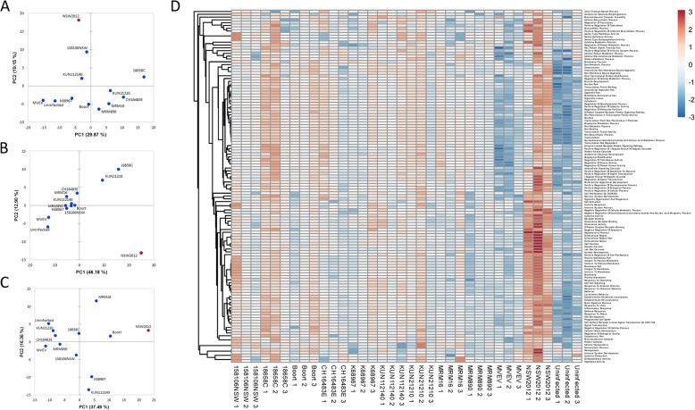
Analysis of WNVKUN induced gene expression in SK-N-SH cells. a-c Principal component analysis plots of gene expression of the infected cells in two dimensions (3 experiments are shown). Isolates are represented as blue dots with the exception of NSW2012 which is represented as a red dot. d Pathway analysis of the triplicate WNVKUN infections. Illustrates pathway score over a > 6 log2 range (> 64-fold) shown arbitrarily on the scale from − 3 to 3. Gene expression is represented in the range from lower (blue) to higher (red)

